
[ad_1]

மரபணுக்களின் வடிவத்தை ஆய்வு செய்வதற்கான நுட்பம் இன்னும் விரிவானது.
புதிய நுட்பம் “ஹப்பிளில் இருந்து ஜேம்ஸ் வெப்பிற்கு மேம்படுத்துவது போன்றது.”
ஒரு புதிய இமேஜிங் நுட்பம் மனித மரபணுவின் முப்பரிமாண கட்டமைப்பை முன்னோடியில்லாத விவரங்களுடன் படம்பிடிக்கிறது, மரபணுவின் முப்பரிமாண கட்டமைப்பை உருவாக்கும் அடிப்படை அலகுகளான நியூக்ளியோசோம்களின் மட்டத்தில் தனிப்பட்ட மரபணுக்கள் எவ்வாறு மடிகின்றன என்பதைக் காட்டுகிறது.
ஜெனோமிக் ஒழுங்குமுறை மையத்தின் (CRG) பார்சிலோனாவைச் சேர்ந்த ஆராய்ச்சியாளர்களால் உருவாக்கப்பட்ட தொழில்நுட்பம் மற்றும் பயோமெடிசின் ஆராய்ச்சி நிறுவனம் (IRB பார்சிலோனா), அதிநவீன கணினி மாடலிங் உடன் உயர் தெளிவுத்திறன் நுண்ணோக்கியை ஒருங்கிணைக்கிறது. மரபணுக்களின் வடிவத்தை ஆய்வு செய்வதற்கு இன்றுவரை இது மிகவும் விரிவான நுட்பமாகும்.
புதிய நுட்பமானது, மரபணுக்களின் முப்பரிமாண மாதிரிகளை உருவாக்கி டிஜிட்டல் முறையில் வழிசெலுத்த ஆராய்ச்சியாளர்களை அனுமதிக்கிறது. மரபணுக்கள் எவ்வாறு செயல்படுகின்றன என்பதைப் புரிந்துகொள்வது, ஆரோக்கியம் மற்றும் நோய் இரண்டிலும் மனித உடலை எவ்வாறு பாதிக்கிறது என்பதைப் புரிந்துகொள்வது, கிட்டத்தட்ட ஒவ்வொரு மனித நோய்க்கும் சில மரபணு அடிப்படைகள் உள்ளன.
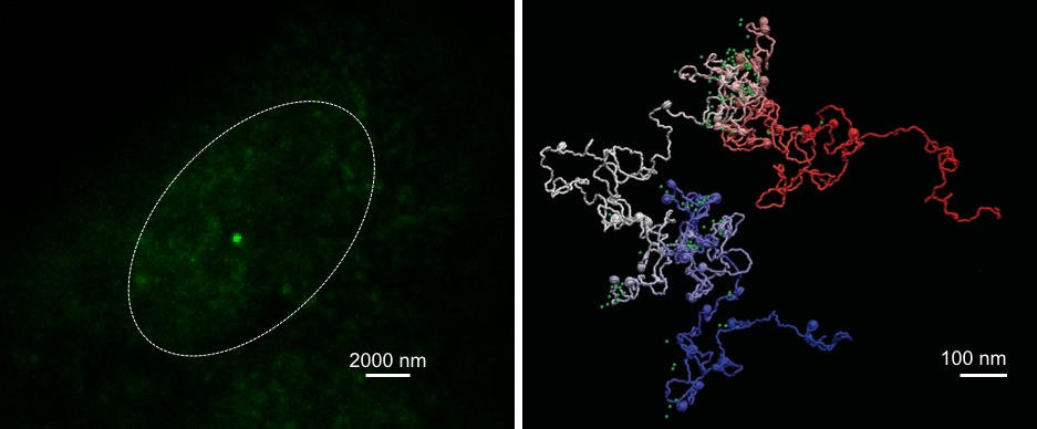
NANOG மரபணுவின் கட்டமைப்பைக் காட்சிப்படுத்த வழக்கமான நுண்ணோக்கியைப் பயன்படுத்தி (இடது) ஒப்பிடுதல், இது தனிப்பட்ட மரபணுக்களைப் படம்பிடிக்கும் MiOS (வலது) ஐப் பயன்படுத்தி பிரகாசமான பச்சைப் புள்ளியாகக் காட்டுகிறது. MiOS சுமார் பத்து மடங்கு தெளிவுத்திறனில் சிறப்பாக உள்ளது மற்றும் வழக்கமான முறைகளைப் பயன்படுத்தி கண்டறிய முடியாத கட்டமைப்பின் முக்கியமான அம்சங்களையும் விவரிக்கிறது. கடன்: Vicky Neguembor/CRG மற்றும் Pablo Dans/IRB பார்சிலோனா
நோயை ஏற்படுத்தும் மரபணுக்களின் கட்டமைப்பில் உள்ள வேறுபாடுகளை பட்டியலிடுவது போன்ற விஷயங்கள் தவறாக நடக்கும்போது மரபணுக்களுக்கு என்ன நடக்கும் என்பதைக் கணிக்க விஞ்ஞானிகள் இறுதியில் இந்த அறிவைப் பயன்படுத்த முடியும். மாறுபட்ட மரபணுவின் வடிவத்தை மாற்றும் மருந்துகளை சோதிக்க இந்த முறை பயன்படுத்தப்படலாம், இது பல்வேறு நோய்களுக்கான புதிய சிகிச்சையின் வளர்ச்சிக்கு உதவுகிறது.
நுண்ணோக்கிகளை உருவாக்குவதன் மூலம் முதன்முதலில் நானூறு ஆண்டுகளுக்கு முன்பு தொடங்கப்பட்ட உயிரினங்களைப் படிக்கப் பயன்படுத்தப்படும் இமேஜிங் நுட்பங்களின் அடுத்த பரிணாமம் தொழில்நுட்பமாகும். மருத்துவம் மற்றும் மனித ஆரோக்கியத்தை மேம்படுத்துவதில் இவை முக்கிய பங்கு வகித்தன, உதாரணமாக, ராபர்ட் ஹூக் முதல் முறையாக செல்களை விவரிக்க பயன்படுத்தினார், பின்னர் நியூரான்களை அடையாளம் காண சாண்டியாகோ ரமோன் ஒய் காஜால் பயன்படுத்தினார். பெரிய முன்னேற்றங்கள் இருந்தபோதிலும், ஆப்டிகல் நுண்ணோக்கிகளின் வரம்புகள் 1873 ஆம் ஆண்டிலேயே தெளிவாக இருந்தன, ஆராய்ச்சியாளர்கள் அவற்றின் அதிகபட்ச தெளிவுத்திறன் 0.2 மைக்ரோமீட்டரைத் தாண்ட முடியாது என்று நிபந்தனை விதித்தனர்.
இந்த இயற்பியல் வரம்பு 21 ஆம் நூற்றாண்டில் சூப்பர்-ரெசல்யூஷன் மைக்ரோஸ்கோபியை உருவாக்குவதன் மூலம் முறியடிக்கப்பட்டது, இது 2014 இல் வேதியியலுக்கான நோபல் பரிசு வழங்கப்பட்டது. ஃப்ளோரசன்ஸைப் பயன்படுத்தி, ஆராய்ச்சியாளர்கள் ஒளி நுண்ணோக்கியின் வரம்புகளை 20 நானோமீட்டர்களில் கைப்பற்றி நிகழ்வுகளை கைப்பற்றினர். முன்னோடியில்லாத மூலக்கூறு அளவில் வாழ்க்கை எவ்வாறு செயல்படுகிறது என்பதை இது வெளிப்படுத்தியது.
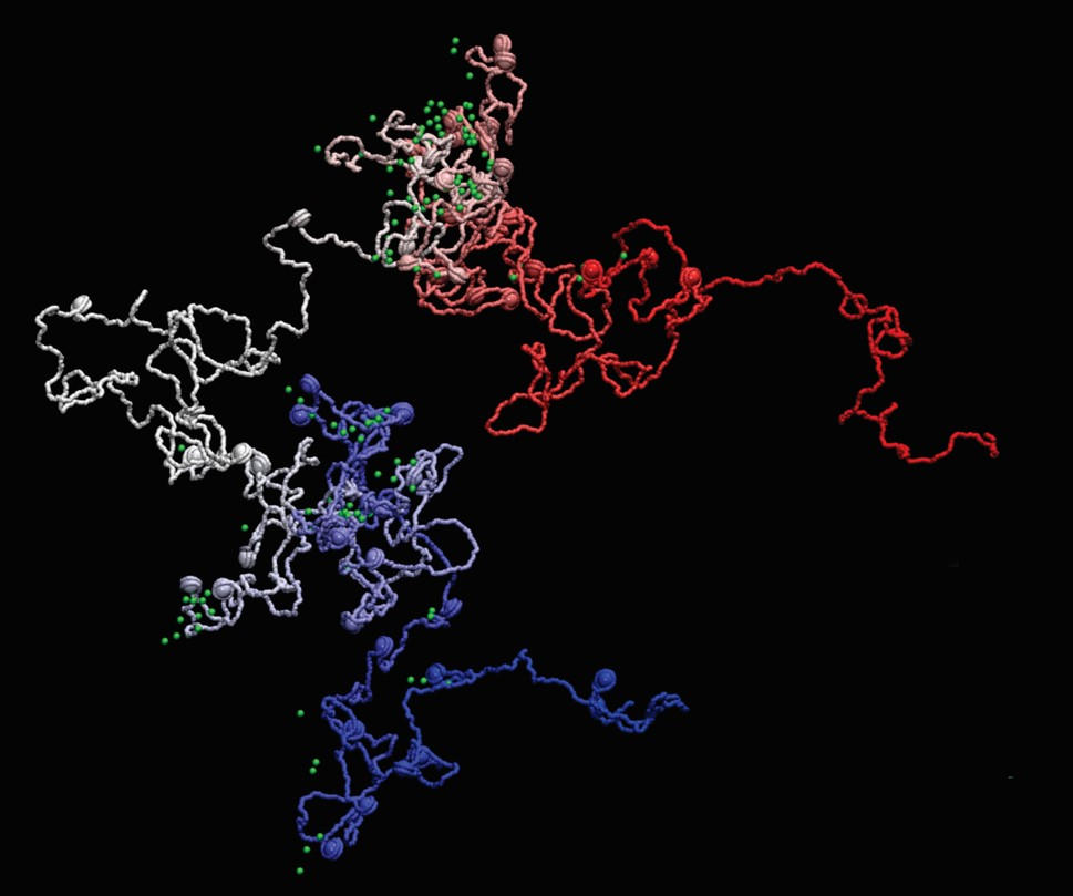
3D இல் மரபணு எவ்வாறு மடிகிறது என்பதைக் காட்டும் MiOS மாதிரியின் எடுத்துக்காட்டு. சில பகுதிகள் எவ்வாறு சுருக்கப்பட்டுள்ளன, மற்றவை நீட்டிக்கப்பட்டு மேலும் அணுகக்கூடியவை என்பதை இது வெளிப்படுத்துகிறது. கடன்: பாப்லோ டான்ஸ்/ஐஆர்பி பார்சிலோனா
சூப்பர்-ரெசல்யூஷன் மைக்ரோஸ்கோபி உயிரியல் மருத்துவ ஆராய்ச்சியின் போக்கை மாற்றியது, விஞ்ஞானிகள் பல்வேறு நோய்களில் புரதங்களைக் கண்காணிக்க உதவுகிறது. இது மரபணு வெளிப்பாட்டைக் கட்டுப்படுத்தும் மூலக்கூறு நிகழ்வுகளைப் படிக்க ஆராய்ச்சியாளர்களுக்கு உதவியது. விஞ்ஞானிகள் இப்போது தொழில்நுட்பத்தை உருவாக்க விரும்புகிறார்கள் மேலும் தகவல்களின் அடுக்குகளைச் சேர்ப்பதன் மூலம் ஒரு படி மேலே செல்ல விரும்புகிறார்கள்.
சூப்பர்-ரெசல்யூஷன் நுண்ணோக்கியை எடுத்து அதை மேம்பட்ட கணக்கீட்டு கருவிகளுடன் இணைப்பது மரபணுக்களை அவற்றின் வடிவம் மற்றும் செயல்பாட்டை ஆய்வு செய்ய தேவையான விவரங்களின் அளவில் இமேஜிங் செய்வதற்கான ஒரு வழியாகும் என்று ஆராய்ச்சியாளர்கள் கருதுகின்றனர். விஞ்ஞானிகளின் ஒரு இடைநிலைக் குழு அவர்களின் நிபுணத்துவத்தைப் பகிர்ந்து கொண்டது மற்றும் மாடலிங் immuno-OligoSTORM – அல்லது சுருக்கமாக MiOS என்ற புதிய நுட்பத்தை உருவாக்கியது.
இரண்டு ஆராய்ச்சி குழுக்களும் பார்சிலோனா இன்ஸ்டிடியூட் ஆஃப் சயின்ஸ் அண்ட் டெக்னாலஜியின் (பிஐஎஸ்டி) இக்னைட் அழைப்பின் ஒரு பகுதியாக இணைந்தது, இது பல்வேறு அறிவியல் துறைகளுக்கு இடையே அறிவு பரிமாற்றத்தை எளிதாக்குகிறது மற்றும் சிக்கலான கேள்விகளுக்கு தீர்வு காண புதிய அணுகுமுறைகளை ஆராய்கிறது.
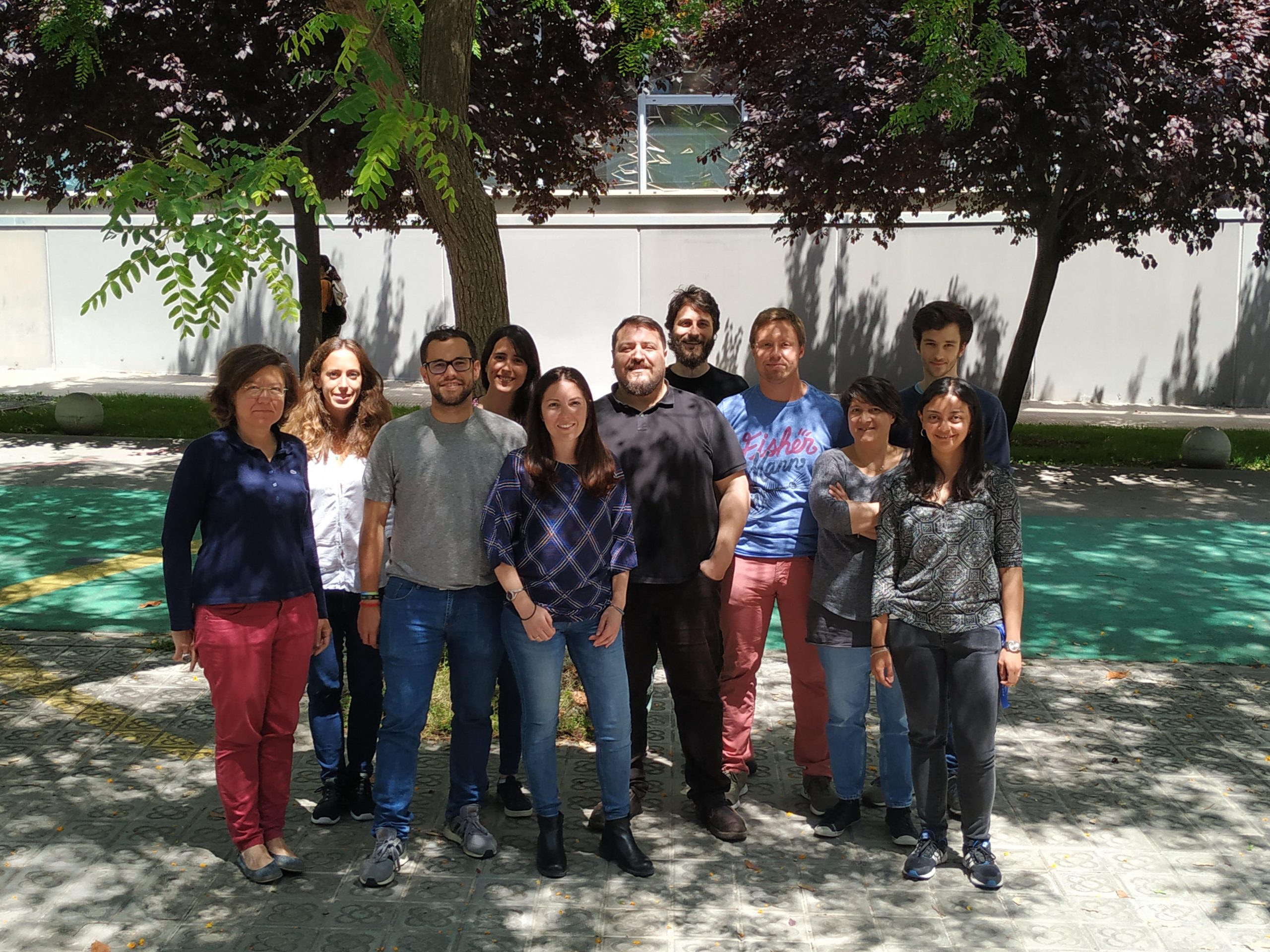
இடமிருந்து வலமாக: Pia Cosma, Laura Martin, Rafael Lema, Ximena Garate, Victoria Neguembor, Pablo Dans, Juan Pablo Arcon, Jürgen Walther, Isabelle Brun Heath, Pablo Romero, Diana Buitrago. கடன்: BIST
“எங்கள் கணக்கீட்டு மாடலிங் உத்தி இதிலிருந்து தரவை ஒருங்கிணைக்கிறது[{” attribute=””>DNA sequencing techniques and super-resolution microscopy to provide an essential picture (or movie) of the 3D shape of genes at resolutions beyond the size of nucleosomes, reaching the scales needed to understand in detail the interaction between chromatin and other cell factors,” says Dr. Juan Pablo Arcon, co-first author of the work and postdoctoral researcher at IRB Barcelona.
As proof of concept, the research team used MiOS to provide new insights on the position, shape, and compaction of key housekeeping and pluripotency genes, revealing new structures and details that are not captured using conventional techniques alone. The findings are published in the journal Nature Structural & Molecular Biology. The study’s corresponding authors include ICREA Research Professor Pia Cosma at the CRG and Professor Modesto Orozco at IRB Barcelona, as well as Pablo Dans, previously a researcher at IRB Barcelona and now at University of the Republic (Uruguay) and the Institut Pasteur of Montevideo.
“We show that MiOS provides unprecedented detail by helping researchers virtually navigate inside genes, revealing how they are organized at a completely new scale. It is like upgrading from the Hubble Space Telescope to the James Webb, but instead of seeing distant stars we’ll be exploring the farthest reaches inside a human nucleus,” says Dr. Vicky Neguembor, co-first and also a co-corresponding author of the study and researcher at the CRG.
While a lot of genome-based research is already changing how we diagnose, treat, or prevent diseases, the impact of MiOS is more long-term. By shedding light on how genes work and how they are regulated at the nanoscale, the technique will enable new discoveries in the scientific laboratory, some of which might eventually translate into clinical practice.
The research team is already putting MiOS to use by exploring genes that are important for human development. The team will also keep developing MiOS further, adding additional functionality that can for example detect how transcription factors – proteins involved in the process of converting or transcribing, DNA into RNA – bind to DNA.
Reference: “MiOS, an integrated imaging and computational strategy to model gene folding with nucleosome resolution” by Maria Victoria Neguembor, Juan Pablo Arcon, Diana Buitrago, Rafael Lema, Jürgen Walther, Ximena Garate, Laura Martin, Pablo Romero, Jumana AlHaj Abed, Marta Gut, Julie Blanc, Melike Lakadamyali, Chao-ting Wu, Isabelle Brun Heath, Modesto Orozco, Pablo D. Dans and Maria Pia Cosma, 11 October 2022, Nature Structural & Molecular Biology.
DOI: 10.1038/s41594-022-00839-y
The study was funded by the Barcelona Institute of Science and Technology, the Horizon 2020 Framework Programme, the Spanish Ministry of Science, Innovation, and Universities, and the Government of Catalonia.
[ad_2]